Perspectives: An Interview with John Newell, MD FACR on Hot Topics in Thoracic Imaging
There is a lot going on right now in the world of thoracic imaging, from coronavirus screening to AI to burnout. To get an insider's perspective on these topics and more, we sat down "virtually" with John Newell, MD FACR for a Q&A.
About John Newell, MD
Dr. Newell has over 30 years of experience as a CardioPulmonary Diagnostic Radiologist working in clinical care, research, administration and education. He was certified in 1980 as a Diagnostic Radiologist by the American Board of Radiology. He is a Fellow of the American College of Radiology, a member of the Fleischner Society, a member of the American Association of Physicists in Medicine and member of IEEE.
Dr. Newell's ongoing professional goals are to improve medical imaging's ability to meet the needs of patients from all countries through improvements in the delivery of clinical imaging, research and education. He specializes in CardioThoracic Diagnostic Radiology, Biomedical Engineering, Medical Physics, and Flight Surgeon.
Our Interview with Dr. Newell
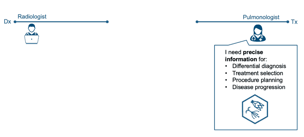
VIDA: VIDA was excited to see COPDGene's recent proposal for a new definition of diagnosing COPD. As one of the authors of that paper, do you have any additional insights to share on how the recommendation was determined or how the inclusion of imaging in diagnosis would impact COPD patients?
Dr. Newell: I think the paper answered the question if COPD should be suspected in the face of normal spirometry and the answer is a resounding "yes" for both positive CT imaging and positive clinical symptoms for COPD as well as expanding the spirometry criteria. Those of us in COPD have suspected this for a long time but proving it required a lot of work.
VIDA: What are the biggest barriers to including Quantitative CT (QCT) imaging routinely as the standard of care for COPD patients?
Dr. Newell: One barrier is in educating radiologists and pulmonologist of the value of quantitative CT in the initial assessment of smokers suspected of having COPD and who are undergoing a non-contrast CT scan or LCS CT scan. I also think advising the operators of CT scanners of the need to use an appropriate CT protocol for the assessment of COPD is an obstacle that needs overcoming.
VIDA: What are your thoughts on CT's role in the diagnosis of 2019-nCoV Coronavirus?
Dr. Newell: Take a look at the recent paper by Bernheim et al which examines early (0-2 days), intermediate (3-5 days) and late (6-12 days) findings on chest CT. The Figures in this paper are very nice and it is the second publication by these authors on COVID-19 pneumonia. 56% of the early disease patients had a normal chest CT scan. The intermediate and late chest CT scans showed predominantly bilateral, peripheral, lower lobe ground glass and consolidative opacities.
VIDA: In the year 2025, how will thoracic radiology be different?
Dr. Newell: I think chest CT studies will routinely be pre-populated with QCT metrics and machine learning (ML) text and the imaging physician will certify the results and serve as an active real time consultant to patients and referring doctors.
Chest CT studies will routinely be pre-populated with QCT metrics and machine learning (ML) text and the imaging physician will certify the results and serve as an active real time consultant to patients and referring doctors."
John Newell, MD
VIDA: As you've explored the market for lung AI applications, including LungPrint by VIDA and others, what types of solutions excite you? How will they add value to your daily workflow?
Dr. Newell: LungPrint Discovery QCT metrics including lung volumes along with the t-MPR images excite me the most in saving me time in the read process and serving as a second reader so I do not miss emphysema or pulmonary fibrosis.
VIDA: We've been hearing radiology leaders urge colleagues to embrace greater clinical collaboration and patient interaction. Do radiologists want this? Are they open to more interaction with patients and referring physicians? Can AI help? Who will drive this effort?
Dr. Newell: The answer to the first 3 questions is "yes."
In reply to your last question--Who will drive this effort?--I hope VIDA is the one leading the case for lung CT AI in both the research and clinical spaces. I think that radiologist's want to increase their efficiency and also increase their value in patient care by embracing AI technologies like VIDA's Lung Print Discovery. VIDA is in a unique position to continue to bring AI applications that are developed by VIDA's research arm and bring these newer AI tools into clinical radiology through Lung Print Discovery. This is I think a win for patients, radiologists and VIDA.
VIDA: Can AI help radiologists overcome everyday challenges and frustrations?
Dr. Newell: It (AI) is a part of the solution. VIDA can bring significant improvements in efficiency for radiologists who read chest CT scans. VIDA can empower the reporting process that the radiologist uses to include valuable AI metrics for the assessment of COPD and ILD and do this in a way that actually increases the efficiency of the radiologist. The workload of radiologists needs to be more realistic and provide for healthy living. The burnout rate among Radiologist is quite high:
From Radiology Business: "An emerging question in radiology is whether radiologists are more susceptible to burnout than physicians in other specialties. Medscape’s latest survey on burnout, released in January and based on responses from more than 15,000 doctors, shows an overall physician burnout rate of 44%. Radiology landed very close to the middle of the pack, at 45%. (Urologists self-reported the most burnout, at 54%, while public health & preventive medicine had the least, 28%.) However, a nationwide study published in Mayo Clinic Proceedings in 2015 showed radiologist burnout at a concerning 61%. Wherever the true figure stands, it’s clear that radiologists face a unique set of stressors. These include turnaround time pressures, ever-increasing imaging volumes, declining reimbursements per study, isolation in the work environment, changes in the market and perceived livelihood threats from technological advances, among others."
The combination of increasing imaging volumes, decreasing reimbursements, isolated work environment and threats of AI technology taking their jobs has increased the stress of being a radiologist. I think VIDA can help in that LungPrint Discovery can increase efficiency as well as provide AI metrics that enhance the value of the radiologists' report and hopefully free up time for the radiologist to interact more with patients, referring physicians and colleagues.
VIDA: What is the biggest threat to thoracic radiologists and pulmonary care in general?
Dr. Newell: Economics. The current system in the US is broken and needs a number of improvements. The cost of drugs, cost of medical equipment, large profit margins for insurers have lead to a difficult economic picture for physicians and other healthcare providers. There has been little pressure to bring the costs of drugs, equipment and insurer profit margins down and so rising healthcare costs due to inflation and new novel but expensive therapies must take that money from patients in the form of cost increases and from healthcare workers in the form of increased workloads with lower compensation rates. The system needs a value based system that is fair to the patient, healthcare worker as well as the healthcare industry. The system needs fair profit margins for all players in the system.
VIDA: What developments in thoracic radiology are most exciting to you?
Dr. Newell: Upright CT scanner from Canon Medical Systems, Photon Counting CT and Generative Adversarial Network (GANs) ML methods.
- Upright CT scanners will be able to assess the lungs in the sitting or standing position which is the same position used in pulmonary function testing. The QCT metrics will more closely align with the global physiologic measures of lung disease.
- Photon counting CT detectors have the potential to greatly reduced the ionizing radiation dose to the patient compared to existing energy integrating CT detectors that have been used since the 1970's in commercial CT scanners. The photon counting systems will also provide more spectral bands for multi-spectral CT scanning.
- GANS may make it possible to predict what an upright CT scan of the lung might look like from a supine CT scan of the lung.
VIDA: Any other thoughts you'd like to share related to trends in thoracic imaging or lung care in general?
Dr. Newell: Early detection of mucus plugs by CT in Asthma and COPD patients will power new therapies for these patients in the near future.
VIDA thanks Dr. Newell for his time and insights in this interview.
-end-